 |
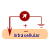 |
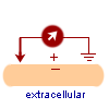
| Figure 3a. Extracellular recording techniques (obtained from [12]). | Figure 3b. Intracellular recording techniques (obtained form [12]). |
In total, 96 direction sensitive neurons were examined:
 |
 |
| Figure 3a. Extracellular recording techniques (obtained from [12]). | Figure 3b. Intracellular recording techniques (obtained form [12]). |
Extracellular techniques are less damaging than intracellular techniques which may actually seriously damage the cell, but may not necessarily be as accurate. Further details regarding these techniques may be found in the references below.
Figure 4 illustrates the actual insertion of the probe at a location
very close to the target cell in area MT.
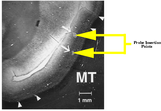 |
| Figure 4. Sample of the actual locations of the insertion of two probes in the cells of area MT. |
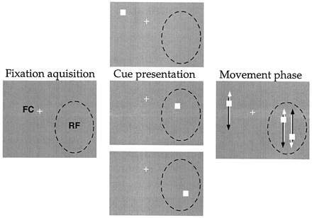
The subject had to then respond to the target by pressing a lever. Pressing, caused distracter dot(s) to appear on the display. In experiment one, one distracter would appear while in experiment two, two distracter dots appeared. The location of the distracter on the display was dependent on the target location. In experiment one, if the target was in the receptive field, then the distracter was not (and vice-versa). In experiment two, when the target was in the receptive field one distracter was not in the receptive field while one was. When the target was not in the receptive field, both distracters were in the receptive field.
In both experiments, when the distracters appeared, all dots including target started moving back and forth on the display: Since the neurons were direction sensitive, one of the directions (up or down) was the neuron's preferred direction of motion. All dots moved with the same speed but possibly in different directions and reversed their direction of motion at the same time. After a random period of time (between 1 - 5s) the speed of the target was increased.
The subject had to respond to this speed increase by releasing the lever which was pressed earlier. If the lever was released within some time interval, the subject was rewarded with apple juice! To complicate matters however, the speed of the distracter dots may have also changed at random times, often before the target's speed was changed. Releasing the lever in this situation caused the trial to terminate without reward. During each trial, the subject's gaze was maintained on the fixation cross and the eye positions were measured every 5ms. The trial was stopped without reward if the subject moved its gaze beyond 1 - 1.5o beyond center of fixation cross.
An illustration of the process described above may be found in figures
5 and 6.
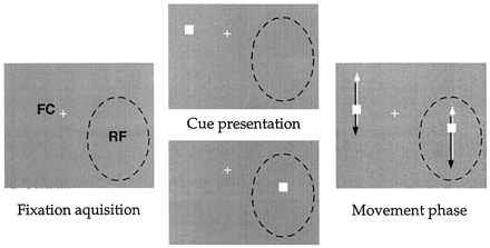 |
| Figure 5. Stimulus and setup of experiment one. |
 |
| Figure 6. Stimulus and setup of experiment two. |